
X Ray Techniques In Dentistry
Emphasis on the use of positioning and placement of the x-ray sensor. The images dental x-rays create help dentists detect abnormalities and diagnose problems such as cavities and root abscesses.

Pin On Dental Assistant Study
About Press Copyright Contact us Creators Advertise Developers Terms Privacy Policy Safety How YouTube works Test new features.

X ray techniques in dentistry. Before the beginning of the 19 th century dentists could only diagnose dental problems by listening to the patients symptoms and looking inside the patients mouths dental. Intraoral Radiographs Bitewing Radiographs Occlusal Radiographs Periapical Radiographs 12. Dental X-ray Techniques II.
Panoramic x-rays help the dentist visualize your entire mouth and have multiple uses including identifying wisdom teeth that need to. An offshoot of dental X-rays is 3D imaging. With this technique the film is placed parallel to the long axis of a tooth allowing the X-ray to be focused perpendicular to the long axis of the tooth.
More years in service decreases the likelihood of applying individual indications for performing a full mouth examination. Ad Find China Manufacturers Of Portable Dental X Ray Unit. Response rate was 53.
Students are given a demonstration of panoramic radiology and gain an understanding of this special procedure. Occlusal x-rays check for underbites or overbites. The patient is seated upright in the dental chair and should remove any removable dental appliances glasses or jewelry that could.
More commonly known as Cone Beam Imaging it creates a panoramic 3D image of a patients entire dental anatomy. The energy of those crashing electrons is converted into a spectrum of x-rays. Only 2 used film technique and solid-state detector SSD was the most used digital technique.
Bitewing x-rays look at how well your back teeth match up on each side and check for cavities between the teeth. Periapical radiographs are probably the most familiar with images of a few teeth at a time captured on small film cards inserted in the mouth. Film development and mounting are discussed and practiced.
Ad Find China Manufacturers Of Portable Dental X Ray Unit. How to Take Bite Wing Dental X-Rays - YouTube. There are several types of extraoral X-rays.
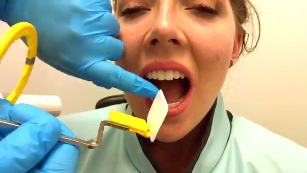
These X-rays are used with low levels of radiation to capture images of the. The patient bites down on a plastic piece that is holding the x-ray sensor and it goes between your back molars and front molars on each side making it a total of four individual x-rays. In simple terms diagnostic x-rays are produced by accelerating electrons across a large voltage and bombarding them onto a heavy metal target usually tungsten.
These important dental x-rays are much simpler to take than a full mouth series of x-rays. Dental X-ray Unit 7. Extraoral X-rays are used to detect dental problems in the jaw and skull.
Recognition of landmarks that may constitute a retake. Extraoral Radiographs OPG DPT Lateral Ceph PA view 2. Orthodontists and dentists use Cone Beam Computerized Tomography CBCT which evolved from CAT Computerized Axial Tomography Scan technology.
The digital dental radiography and x-ray course is intended to be a brush up on the placement and principles of the digital sensor technique that can be used in clinical settings. This occurs inside an x-ray tube. Dental x-rays help dentists see what is happening to parts of the mouth that he or she cannot visualize with the naked eye or with the help of dental tools.
X-ray technique Registered Dental Hygienists. Digital radiography is a type of X-ray imaging that uses digital X-ray sensors to replace traditional photographic X-ray film producing enhanced computer images of teeth gums and other oral structures and conditions. X-ray Radiography There are three types of diagnostic radiographs taken in todays dental offices -- periapical also known as intraoral or wall-mounted panoramic and cephalometric.
Students learn how to expose dental periapical x-ray film using the Rinn Paralleling Technique using the XCP film holder. Ninety-eight percent of the dentists had made the transition to digital radiography. Occlusal X-rays track the development and placement of an entire arch of teeth in either the upper or lower jaw.
Dental X-rays radiographs are images of your teeth that your dentist uses to evaluate your oral health. X-rays and How They Are Produced. Dental professionals today are increasingly using digital dental radiographs digital X-rays to better detect diagnose treat and monitor oral conditions and diseases.
DC is a high frequency intraoral X-Ray generator used to obtain radiographs of the oral cavity that allow dentists to see inside a tooth and beneath the gums to assess.

Veterinary Dental Radiography Techniques Vet Tech School Veterinary Tech Vet Medicine

Pin By Megan On X Ray Radiology Student Radiology Technician Radiology Schools
Dental Radiographs Vet Tech School Vet Medicine Vet Tech Student

Technique Charts Where Was This When I Had To Make One Rad Tech Humor Radiology Schools Radiology Student

Radiologic Technique Charts Tech Nique Chart For Techniquetechnique Charts Radiologic Technology X Ray Radiology Radiology Technician

X Ray Using Parallel Cone Technique X Ray Dental Techniques

Digital X Ray Technique Chart Technique Charts Radiology Student Medical Radiography Radiology Schools

Dental X Ray Radiation Safety Comparison Chart Dental X Ray Dental Facts

Dental Hygiene Radiology Processing And Technique Errors Dental Hygienist School Dental Assistant Study Dental Hygiene School
How To Take Periapical Radiographs Dental Health Kindergarten Dental Assistant Study Dental Assistant

X Ray Technique Chart Google Search Xray Technician Radiology Student Radiography Student

Bisecting Angle Technique Vet Medicine Vet Tech Student Dental Hygiene School

Dental X Rays Bitewing X Rays Pdf Free Pdf Epub Medical Books Dental Hygiene Student Dental Hygiene Instrument Dental Hygiene School

Dental X Ray Tube Dental Exam Dental Dental Hygiene Student
Posting Komentar untuk "X Ray Techniques In Dentistry"