
F-techniques Microscopy
The specimen is normally placed close to the microscopic lens. Modern Research and Educational Topics in Microscopy.
Confocal Microscopy Principle Applications Ibidi
Try Thermo Fisher Cell Counter.

F-techniques microscopy. December 01 - 02 2021 Duration. C3 PLO 4 CTPS2 MQF LOD 6 Learning Outcomes. Microscopy was a great breakthrough in the field of nanotechnology.
These techniques include FRET fluorescence resonance energy transfer FLIM fluorescence lifetime imaging microscopy FCS fluorescence correlation spectroscopy and FRAP fluorescence recovery after. In compound microscopes the actual magnification is calculated as the magnification of objective lens multiplied by the magnification power of the ocular lens. Standard detail exposed by the fracture plane is then seen by vacuum deposition of platinum-carbon to make a replica for examination by TEM.
Gerd Binnig and Dr. Preparatory Techniques in Transmission Electron Microscopy. Fundamental knowledge about confocal laser scanning microscopy.
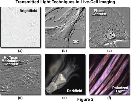
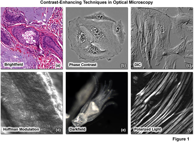
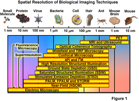
Stimulated emission depletion STED Saturated structured illumination microscopy SSIM REversible Saturable Optical Linear Fluorescence Transitions RESOLFT Photoactivated localization microscopy PALM Fluorescence photoactivation localization microscopy FPALM. High speed wired or wireless internet connection. These include bright field dark field oblique illumination fluorescence phase contrast confocal deconvolution differential interference contrast and dispersion staining microscopy to name a few.
Techniques Uses Fixation Glutaraldehyde osmium tetroxide Standard fixation and postfixation techniques for TEM Paraformaldehyde Less destructive than glutaraldehyde on proteins. Optical microscopy has several drawbacks. In such cases depending on the problem routine diagnostics may be supplemented with specific microscopic techniques including electron microscopy laser scanner microscopy and laser microdissection techniques in order to isolate single cells or cell groups.
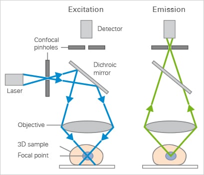
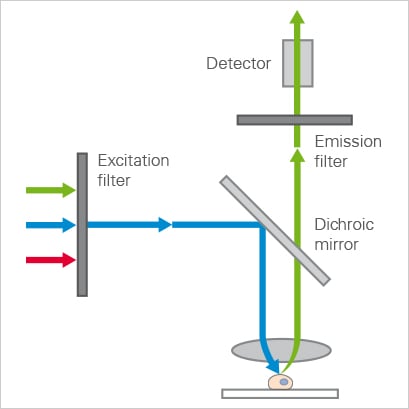
Firstly the technique works at its best only with darker objects or ones that refract effectively. Confocal microscopy is an optical imaging technique that provides very high spatial resolution and contrast compared with the conventional wide-field optical microscopy with additional advantages such as control over field depth minimal background signature and ability to collect serial optical sections from thick specimens 24. Freeze-fracture electron microscopy FFEM is a technique for examining the ultrastructure of rapidly frozen biological samples by TEM.
This practical manual provides an introduction to the so-called F-techniques and their use in probing protein structure and function by leading scientists in fluorescence microscopy. Try Thermo Fisher Cell Counter. To handle microscope properly ii.
Theory of other Photomanipulation strategies eg. Our lntroduction to Confocal Laser Scanning Microscopy Date. At the end of this lesson students should be able to.
FFEM consists of physically breaking apart fracturing a frozen biological sample. They are used however when specific problems occur. Your Time is too Important to Keep Counting Cells Manually.
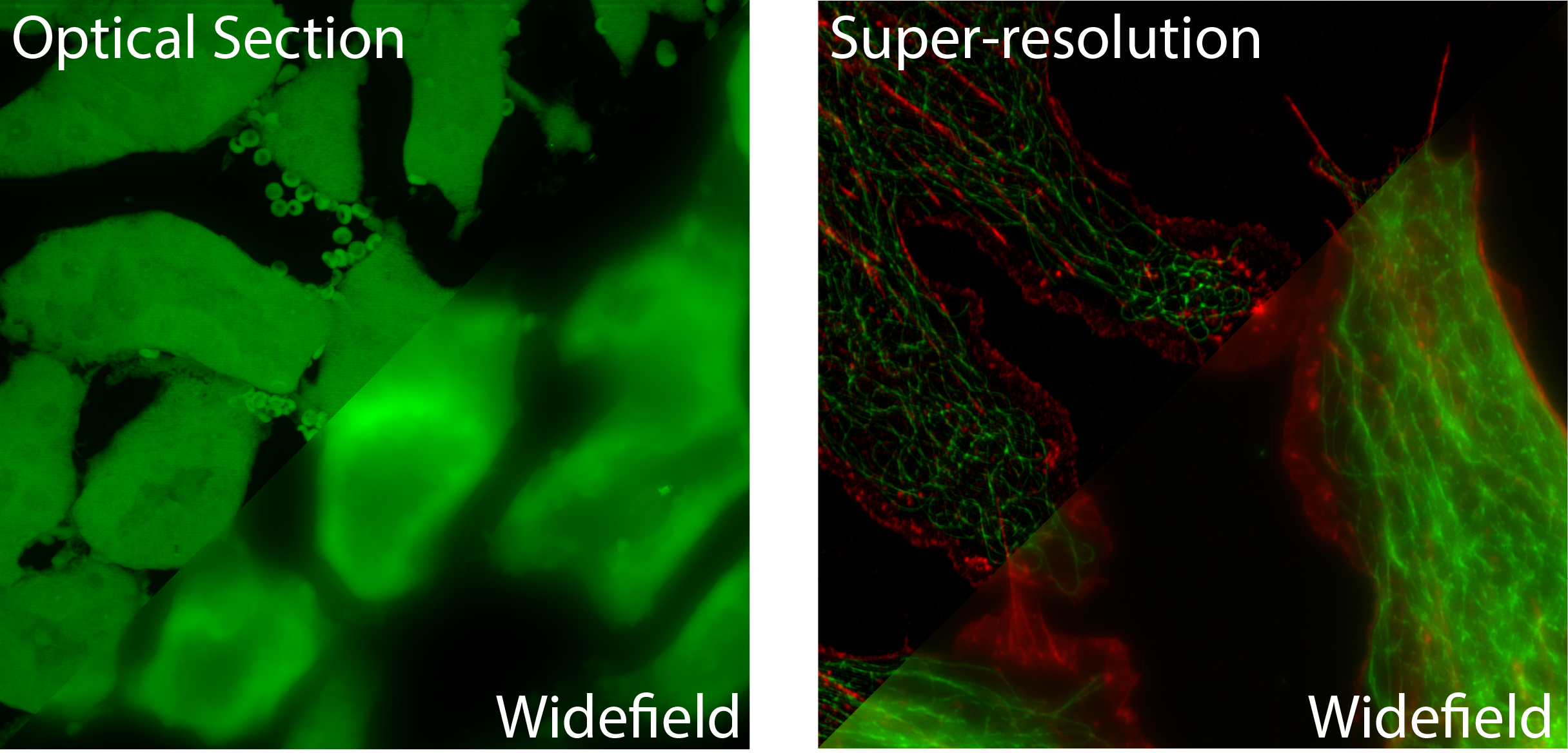
Confocal microscopy offers several advantages over conventional widefield optical microscopy including the ability to control depth of field elimination or reduction of background information away from the focal plane that leads to image degradation and the capability to collect serial optical sections from thick specimens. Advances in the field of fluorescent labelling eg fluorescent proteins quantum dots tetracystein domains and optics eg super-resolution techniques and quantitative methods not only provide better images of the cytoskeleton but also offer an opportunity. Several improvements in microscopy techniques have been invented in the 20th century and have resulted in increased resolution and contrast to some extent.
They use lenses to focus light on the specimen magnifying it thus producing an image. To obtain accurate images. FRET Forster Resonance Energy Transfer a technology to prove molecular interaction on the basis of confocal light microscopy.
Solve basic problems related to cells biomolecules inheritance genetics and biological development. Ad Cell Counting Viability Apoptosis and Transfection Efficiency in Less Than 10 Seconds. The multiple imaging modes afforded by widefield confocal and multiphoton fluorescence microscopies permit noninvasive temporally resolved imaging of fixed and living cells and tissues with a high level of biochemical specificity.
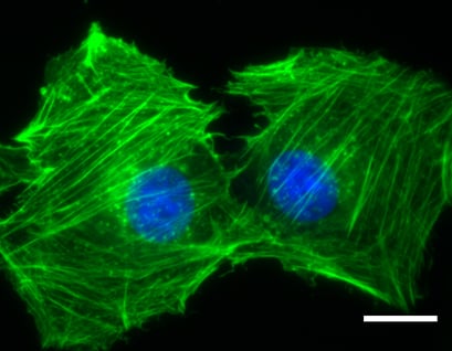
The microscopy techniques used for polymer biomaterials include light microscopy optical and stereo scanning electron microscopy SEM transmission electron microscopy TEM and scanning probe microscopies including atomic force microscopy AFM. Despite the advantages of traditional fluorescence microscopies the spatial resolution of such techniques is. Fluorescence microscopy is a non-invasive technique that allows high resolution imaging of cytoskeletal structures.
However they did not overcome the diffraction limit. Ad Cell Counting Viability Apoptosis and Transfection Efficiency in Less Than 10 Seconds. Fluorescence microscopy is central to many techniques which aim to reach past this limit by specialized optical configurations.
Díaz Eds _____ Microscopy techniques and the study of synapses Emma Perez-Costas Miguel Melendez-Ferro and Rosalinda C. Theory of other Photomanipulation strategies eg. Now that scientists could see individual atoms they could begin to manipulate these atoms to build different structures and study their properties.
Immunocytochemistry Microwave acceleration Short treatment at low-power microwave decreases fixation times21. There are deterministic and stochastic functional techniques. Your Time is too Important to Keep Counting Cells Manually.
The image that seen under microscopes was inverted because it goes through two lens systems and because of the reflection of light rays. A light microscope is a biology laboratory instrument or tool that uses visible light to detect and magnify very small objects and enlarging them. Fundamentals are recommended eg.
BASIC TECHNIQUES IN MICROSCOPY Course Learning Outcome. FRET Forster Resonance Energy Transfer a technology to prove molecular interaction on the basis of confocal light microscopy. Roberts Department of Psychiatry and Behavioral Neurobiology University of Alabama at Birmingham Sparks Center 1720 7th Avenue South Birmingham Alabama USA Microscope.
Confocal Microscopy Principle Applications Ibidi
Zeiss Microscopy Online Campus Live Cell Imaging Microscopy Techniques
Zeiss Microscopy Online Campus Superresolution Structured Illumination Microscopy


How To Prepare Your Specimen For Immunofluorescence Microscopy Science Lab Leica Microsystems

Super Resolved Live Cell Imaging Using Random Illumination Microscopy Sciencedirect
Zeiss Microscopy Online Campus Microscopy Basics Enhancing Contrast In Transmitted Light
Widefield Fluorescence Microscopy Principle Applications Ibidi
Widefield Fluorescence Microscopy Principle Applications Ibidi
Which Fluorescence Microscopy Techniques Is Best For Me
Zeiss Microscopy Online Campus Introduction To Superresolution Microscopy

An Introduction To The Light Microscope Light Microscopy Techniques And Applications Technology Networks

Epifluorescence Microscopy An Overview Sciencedirect Topics
Olympus Fluoview Resource Center Theory Of Confocal Microscopy

Zeiss Microscopy Online Campus Live Cell Imaging Microscopy Techniques
Posting Komentar untuk "F-techniques Microscopy"