
G Banding Techniques Ppt
G-banding G banding or Giemsa banding is a technique used in cytogenetics to produce a visible karyotype by staining condensed chromosomes. G - banding.
Chromosome Banding And Painting Online Biology Notes
The most common R-bandingmethodinvolvesheatdenaturingchromosomesin.

G banding techniques ppt. Most G-banding techniques require pretreating the chromosomes with a proteolytic enzyme such as trypsin. Chromosomal Region Banding Technique Constitutive heterochromatin C-banding Facultative heterochromatin G- or Q-banding Euchromatin R-banding. G-banding most commonly is introduced by treatment with a proteolytic enzyme such as trypsin followed by staining with Giemsa which binds DNA.
We will discuss G-banding in the most detail because you will likely see G-banding. These produce a multicoloured banding pattern that like G-banding is unique for each chromosome Figure 2d. Moreover GTG banding is most popular among all methods.
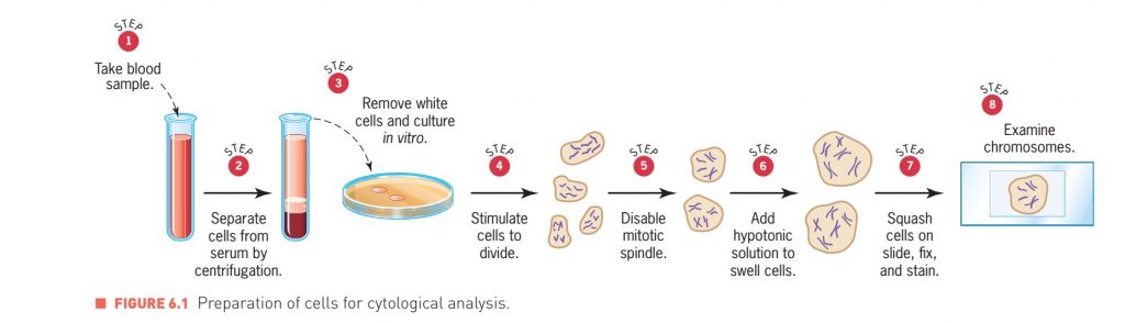
Chromosome banding techniques and staining Giemsa has become the most commonly used stain in cytogenetic analysis. GTG banding Quinacrine banding C-banding and R-bandings are different popular techniques employed to discover various aberrations. White blood cells Fixation Slide preparation MOLECULAR BIOLOGY Overview of.
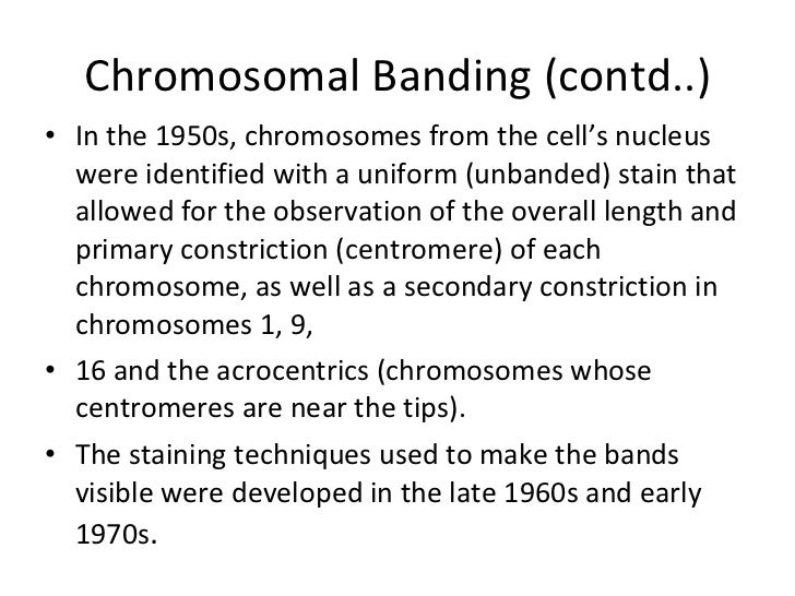
Also called Giemsa chromosome banding. G banding chromosomes - explains about the karyotyping proces. These landmarks facilitate assessment of chromosome normalcy identification of sites.
G-banding gives you a series of light and dark stripes along the length of the chromosome. Chromosomal regions and banding techniques that reveal them. Chromosome G banding is usually performed either by the usage of trypsin which removes chromosomal proteins or by incubation in hot.
The chromosomes are visualized as consisting of a continuous series of bright and dark bands. 15 metaphases are analysed but in specific cases even 50 to 100. Metaphase spread Chromosome Sister Chromatides Metaphase chromosomes Karyotyped chromosomes Banding patterns on human mitotic chomosomes due to regions of condensed chomatin darker - G bands and less condensed chromatin lighter - R bands human chromosome 4 at varying resolutions due to exact mitotic stage or degrees of spreading - squashing - stretching Human.
Each of these techniques provides a useful tool for evaluating complex chromosomal abnormalities in humans for rapidly constructing karyotypes for other species and for performing comparative genome mapping. Basis for G-R-banding G-banding involves staining protease-treated chromo-somes with Giemsa dye and is thought to result from interactions of both DNA and protein with the thiazine and eosin components of the stain. Culturing Slide Making and G Banding I.
An analysis of metaphases with the banding resolution of 400-550 is used and in selected cases metaphases are analysed with high resolution HRT of 700 bands. G-banding involves stai ning p rotease- treated chromosom es with G iemsa dye and is thought to result from in teractions of. Human Chromosomes Identification by G-Banding - Human Chromosomes Identification by G.
The banding techniques fall into two principal groups. R-banding technique also uses Giemsa stain but before staining with Giemsa the slide is. G-banding is unique for each chromosome Figure 2d.
SKY1 is a registered trademark of Applied Spectral. G-banding produces a banding pattern that can be correlated with Q-banding with the G-light bands equivalent to the Q-dull regions and the G-dark bands equivalent to the brightly fluorescent regions. It is useful for identifying genetic diseases through the photographic representation of the entire chromosome complement.
Each of these techniques provides a useful tool for evaluating complex chromosomal abnormalities in hu-mansforrapidlyconstructingkaryotypesforotherspecies andforperformingcomparativegenomemappingNote. With this banding technique the band pattern produced in chromosome is reversed to the band produced by G-banding and Q-banding. In this video we discuss about theChromosome banding technique types G banding karyotyping.
As a standard G-banding technique Giemsa-Trypsin-Wrights is used and in selected cases C-banding AgNOR and FISH techniques. G banding Giemsa Banding Giemsa Banding is the most frequently used banding technique in cytogenetic laboratories. Giemsa-Trypsin-Giemsa banding is implemented to know numerical and structural chromosomal variations.
The common banding techniques that reveal these various chromosomal regions are summarized in table 31. G - banding G bands may be defined as a system of dark and light bands throught the length of the euchromatin part of the chromosomes. The dark band AT rich region observed in G-banding technique appears light in R-banding and vice versa.
A staining technique in which chromosomes are treated with trypsin then with Giemsa stain. 1 those resulting in bands distributed along the length of the whole chromosome such as G- Q- and R-bands and 2 those that stain a restricted number of specific bands or structures. 2G Banding Techniques Chromosome Weak Trypsin urea protease Treated with Giemsa Banding pattern To denature protein Interaction of the DNA with thiazine eosin components of stain brightens sulphur rich regions Methylene Azure Methylene Violet Methylene Blue Eosine.
G-banding preferentially stains the. PowerPoint PPT presentation free to view. This kind of analysis is used in prenatal.
G-Banding Technique Giemsa is the most commonly used stain in cytogenetic analysis Staining a metaphase chromosome with a Giemsa stain is referred to as G-banding Unlike Q-banding most G-banding techniques require pretreating Chromosomes are pretreated with either salt or a proteolytic protein-digesting enzyme. INTRODUCTION During metaphase of the cell cycle the condensed chromosomes align along the metaphase plate prior to chromatid separation and then move along the spindle fibres to the poles of the spindle. The standard is banding methods most often the GTG technique G-banding method using trypsin for selective denaturation of protein structures and dye blocking that yields chromosome-specific.
Chromosome banding techniques produce a series of consistent landmarks along the length of metaphase chromosomes that allow for both recognition of individual chromosomes within a genome and identification of specific segments of individual chromosomes.

Banding Techniques Ppt Copy Ppt Cytogenetics Is The Study Of The Structure And Properties Of Chromosomes Chromosomal Behaviour During Mitosis And Course Hero

Prime Lending Bill Pay Primelending Loanadministration Com Paying Bills Bills Paying

Head Of Marketing Resume Example Cv Template Director Manager Digital You Can Get The Fully Editabl Marketing Resume Resume Skills Project Manager Resume
Karyotyping
Karyotyping G Banded Service

I2c Lcd With Arduino Wiring Diagram Schematic Pinout Arduino Arduino Circuit Lcd

Diagnostics Free Full Text Prenatal Diagnosis Of True Fetal Mosaicism With Small Supernumerary Marker Chromosome Derived From Chromosome 16 By Funipuncture And Molecular Cytogenetics Including Chromosome Microarray
Karyotyping

Chromosome Banding Techniques Youtube

G Banding An Overview Sciencedirect Topics
Karyotyping
Content Strategy For Google Plus Infographic Infographic Marketing Strategy Infographic Social Media Infographic
Human Karyotype Chromosomes

Chromosome Banding Techniques G Banding Karyotyping Techniques Vinay Rajput Youtube
Posting Komentar untuk "G Banding Techniques Ppt"