
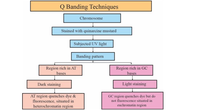
Q Banding Techniques
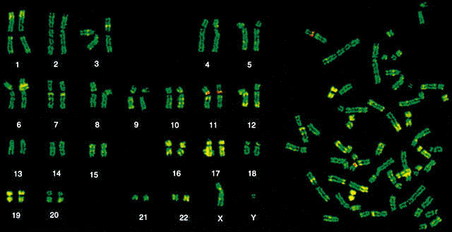
They can be recognized by a yellow fluorescence of differing intensity. The need to combine chromosome banding with fluorescence in situ hybridization has meant that banding techniques using fluorescent dyes has become more popular.
Learn About Banding Pattern Chegg Com
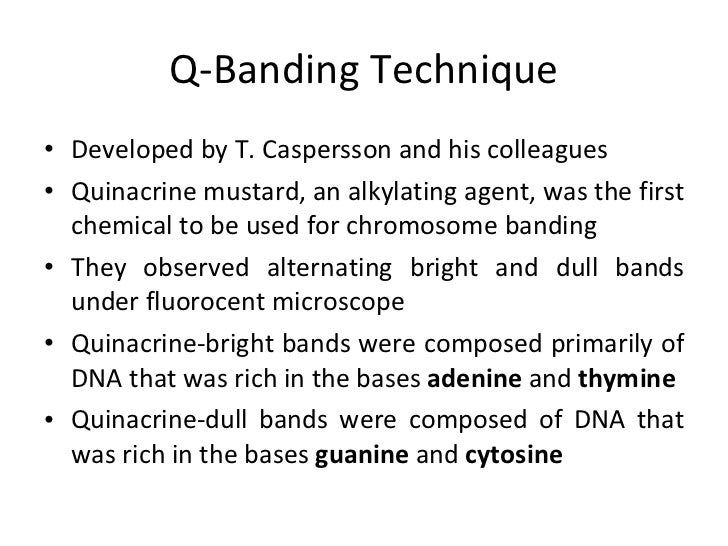
Q banding used quinacrine stain quinacrine dihydrochloride or quinacrine mustard and it is the simplest and the first chromosomal banding method.

Q banding techniques. C-banding only stains the centromeres. And region 1 band 7 sub band 6 respectively. Redirected from Q-banding Q-banding stain.
A fluorescent stain for chromosomes that produces specific banding patterns for each pair of homologous chromosomes. Specific staining protocols include C-banding and staining of the nucleolar organizing regions NORs using a silver stain Ag-NOR. The combined techniques allow more precise identification of marker chromosomes and more accurate definition of breakpoints in the consistent chromosome translocations that are associated with certain human malignancies.
Most part of the stained DNA is heterochromatin. By an DNA intercalating agent quinacridine of metaphase chromosomes. There are several types of chromosomal banding techniques.
G-Banding although Q-Banding also will produce darker regions that are AT-rich Female patient with a translocation between the p and q arms of chromosomes 1 and 14 and region 2 band 1 sub band 3. Q-banding is a fluorescent pattern obtained using quinacrine for staining. Quinacrine banding is thought to reflect the distribution of rnoooooooooooooooo.
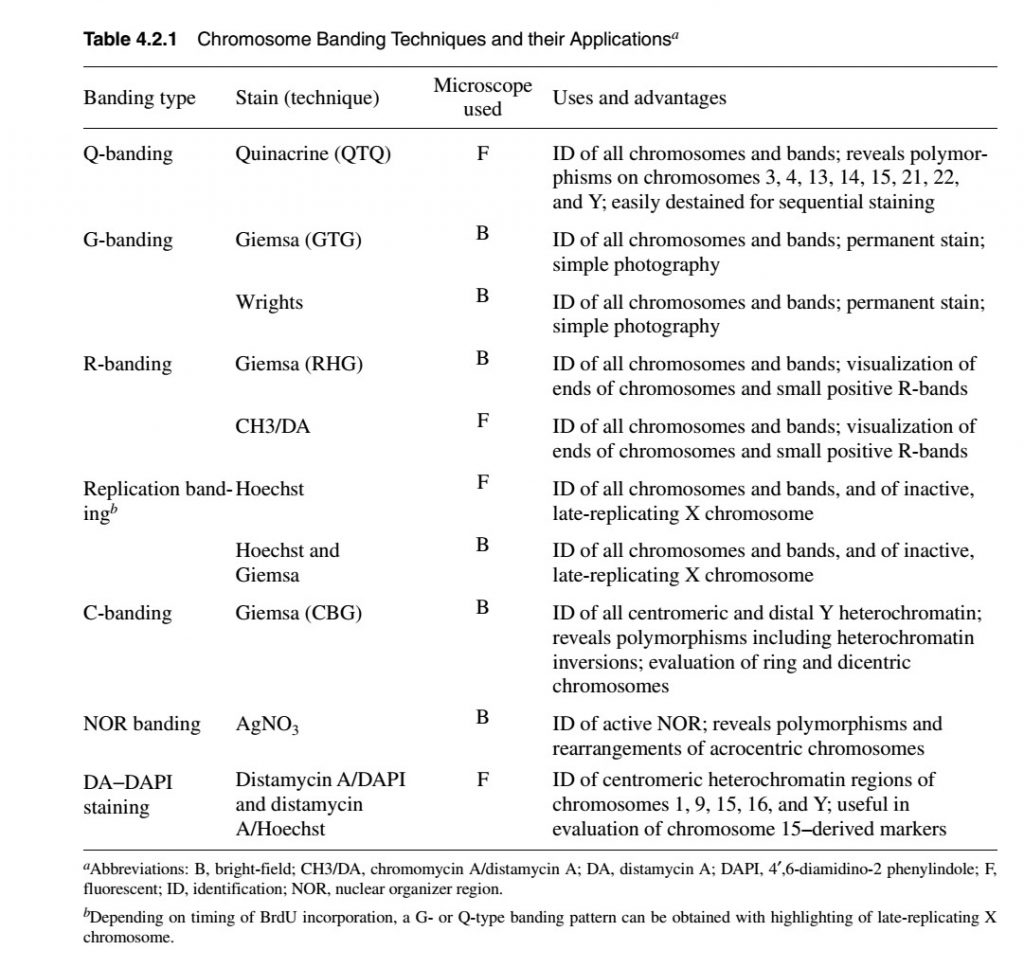
The common banding techniques that reveal these various chromosomal regions are summarized in table 31. The Metaphase Chromosomes are stained in order to visualise the Chromosomes. Q-banding uses a stain called quinacrine.
The pattern of bands is very similar to that seen in G-banding. Centromeric regions of human chromosomes 3 4 and 13 are specifically stained as are satellites of some acrocentric chromosomes and the end of the long axon of the Y. A procedure for the sequential staining of metaphase cells to provide R-banding Q-banding and C-banding of chromosomes is described.
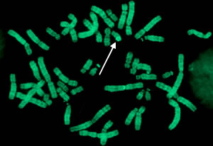
Constitutive heterochromatin nucleolar organizer regions and telomere regions C Ag-I. Fluorescent Q-banding is produced simply as a result of staining with no other treatment necessary. INTRODUCTION-A chromosome banding pattern is comprised of alternating light and dark stripes or bands that appear along its length after being stained with a dyeOR The treatment of chromosomes to reveal characteristic patterns of horizontal bands like bar codes is known as chromosomal banding.
1Q Banding Techniques Chromosome Stained with Quinarcine Mustard Subjected UV light Banding Pattern Region rich in GC bases Region Rich in AT bases Light stainingDark staining GC region quenches dye but do not fluorescence situated in euchromatin region AT region quenches dye fluorescence situated in heterochromatin region 22. Q-banding is technically among the simplest banding technique. It is similar in pattern to G-banding but glows yellow.
Q banding Quinacridine Banding Was the first banding technique discovered Caspersson et al 1969 and up to date it remains the simplest. The banding techniques fall into two principal groups. The Chromosome is a staining technique for chromosomes.
Q banding uses fluorescent staining ie. The chromosomes are stai. Chromosomal Region Banding Technique Constitutive heterochromatin C-banding Facultative heterochromatin G- or Q-banding Euchromatin R-banding.
Bands distributed along the length of the whole chromosome such as G- Q-band Those that stain a number of specific bandsThese include methods which reveal centromeric bands C-bands and nucleolus organizer regions NORs at terminal regions of chromosomes. Chromosomal regions and banding techniques that reveal them. The development of this fluorescent banding technique and its appli-cation to human chromosomes have revealed extensive longitudinal differentiation of chromosomes.
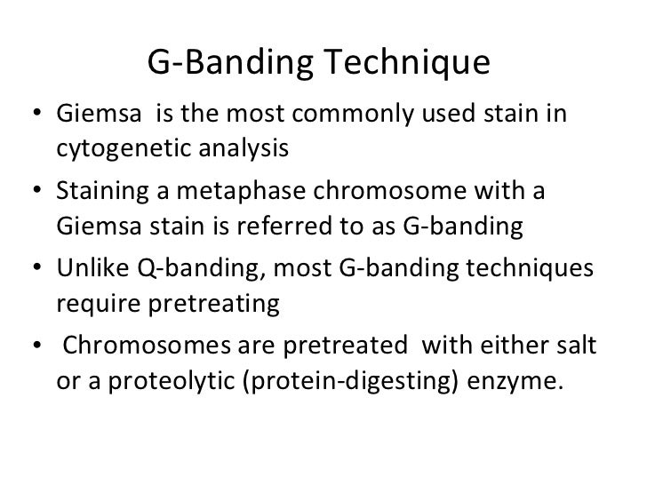
In 1968 the description of the first heterogeneous banding technique of chromosome based on staining with quinacrine dihy-drochloride Q-banding was a revolution on cytogenetics. The staining of chromosome is known as banding technique because stains give rise to pattern of bands along the length of chromosome. G banding technique uses a stain called Giemsa stain and it stains AT-rich regions of heterochromatic regions in darkly stained bands and GC rich euchromatic regions in lightly stained bands.
The Q banding or quinacrine banding technique in which the fluorescence dye quinacrine binds or intercalates with the DNA and gives different banding patterns on chromosomes The technique is also known as QFQ banding- Q-bands using fluorescence quinacrine and was first explained by Caspersson and co-workers. Types of chromosome banding technique. This unit covers these basic banding techniques Q-banding G-banding and R-banding which produce virtually identical patterns of bands along the length of human chromosomes although the bands and polymorphic regions highlighted may differ with each technique.
The basic banding methods are complemented by techniques that stain selective chromosome regions. A unique banding pattern. Among them Q-banding Reverse R banding and G-banding are generalized banding techniques.
The Q-banding name is given by the use of quinacrine fluorescent dye in the banding technique. Generalized banding techniques have included Q-banding reverse banding and G-banding 123031. The present method is similar to the G-banding however here the fluorescent dye is utilized instead of a normal dye.
Q-banding involves staining with quinacrine. ThanR-bandingItisthoughttoidentifytheGC-richestR-bands of which approximately half occur at telomeres in the human genomehencethe name. Q-banding yields a fluorescent pattern.
This unit covers these basic banding techniques Q-banding G-banding and R-banding which produce virtually identical patterns of bands along the length of human chromosomes although the bands and polymorphic regions highlighted may differ with each technique.

Chromosomal Banding Techniques Ppt Video Online Download
Q Banding
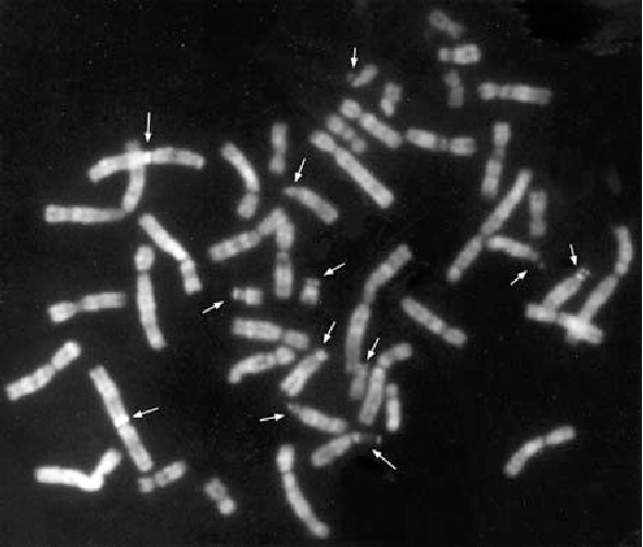
Table 2 2 From 2 1 Chromosome Banding Techniques And Mechanisms 2 1 1 Q Banding Semantic Scholar

Table 2 2 From 2 1 Chromosome Banding Techniques And Mechanisms 2 1 1 Q Banding Semantic Scholar
2
Routine Cytogenetic Analysis Cytogenetics Core Mayo Clinic Research
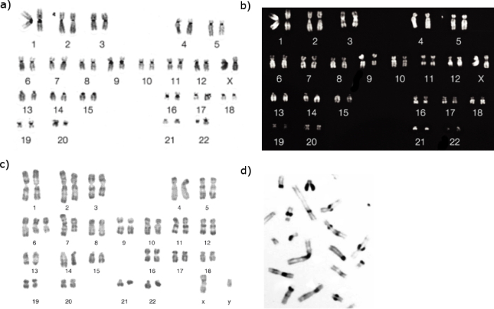
Karyotyping

Neet Ug Bonus Lesson Important Chromosome Banding Techniques Offered By Unacademy
Chromosome Banding As Revealed By Different Staining Techniques Learn Science At Scitable
Karyotyping

Chromosome Banding And Mechanism Of Chromosome Aberrations Intechopen

Chromosome Banding Introduction The Earliest Techniques Stained Chromosomes
Uses Of Banding Techniques For The Identification Of Human Diseases Of Cytogenetic Origin Semantic Scholar
Chromosome Banding And Painting Online Biology Notes
Posting Komentar untuk "Q Banding Techniques"